Chapter Review
Key Terms
Terms in bold are defined in the glossary.
- template
- semiconservative replication
- replication fork
- origin
- Okazaki fragment
- leading strand
- lagging strand
- nucleases
- exonucleases
- endonucleases
- DNA polymerases
- DNA polymerase I
- primer
- primer terminus
- processivity
- proofreading
- DNA polymerase III
- replisome
- helicases
- topoisomerases
- primases
- DNA ligases
- DNA unwinding element (DUE)
- AAA+ ATPases
- catenane
- prereplicative complex (pre-RC)
- licensing
- minichromosome maintenance (MCM) protein
- ORC (origin recognition complex)
- DNA polymerase ε
- DNA polymerase δ
- DNA polymerase α
- mutation
- DNA glycosylases
- base-excision repair
- AP site
- abasic site
- AP endonucleases
- DNA photolyases
- error-prone translesion DNA synthesis
- SOS response
- homologous genetic recombination
- site-specific recombination
- DNA transposition
- recombinational DNA repair
- branch migration
- Holliday intermediate
- replication restart primosome
- meiosis
- double-strand break repair model
- nonhomologous end joining (NHEJ)
- transposon
- transposition
- insertion sequence
Problems
1. DNA Replication An investigator adds DNA polymerase III holoenzyme, DNA primase (DnaG), single-stranded DNA–binding protein (SSB), an ATP-dependent DNA ligase, and the replicative helicase (DnaB) to each of the DNA substrates shown, along with ATP and all four dNTPs. The length of DNA in the circle in structure 1 is 10,000 bp. The linear (unbranched) part of structure 2 is 20,000 bp long.

- If precursor (dNTP) concentrations are not limiting, Okazaki fragments are 2,000 nucleotides long, and replication proceeds at 1,000 nucleotides/s for 30 seconds, which DNA substrate will generate a longer replication product?
- For structure 1, draw the product of this 30 second replication.
- Draw the expected product if DnaG were left out of the reaction.
- Draw the expected product if DNA ligase were instead left out of the reaction.
2. Heavy Isotope Analysis of DNA Replication A researcher switches a culture of E. coli growing in a medium containing to a medium containing for three generations (an eightfold increase in population). What is the molar ratio of hybrid DNA to light DNA at this point?
3. Replication of the E. coli Chromosome The E. coli chromosome contains 4,641,652 bp.
- How many turns of the double helix must be unwound during replication of the E. coli chromosome?
- Using the data in this chapter, how long would it take to replicate the E. coli chromosome at 37 °C if two replication forks proceeded from the origin? Assume replication occurs at a rate of 1,000 bp/s. Under some conditions, E. coli cells can divide every 20 min. How might this be possible?
- In the replication of the E. coli chromosome, about how many Okazaki fragments would be formed? What factors are required to link together newly synthesized Okazaki fragments in the lagging strand?
4. Base Composition of DNAs Made from Single-Stranded Templates Predict the base composition of the total DNA synthesized by DNA polymerase on templates provided by an equimolar mixture of the two complementary strands of bacteriophage DNA (a circular DNA molecule). The base composition of one strand is A, 24.7%; G, 24.1%; C, 18.5%; and T, 32.7%. What assumption is necessary to answer this problem?
5. DNA Replication Kornberg and his colleagues incubated soluble extracts of E. coli with a mixture of dATP, dTTP, dGTP, and dCTP, all labeled with in the α-phosphate group. After a time, they treated the incubation mixture with trichloroacetic acid, which precipitates the DNA but not the nucleotide precursors. They then collected the precipitate and determined the extent of precursor incorporation into DNA from the amount of radioactivity present in the precipitate.
- If any one of the four nucleotide precursors were omitted from the incubation mixture, would radioactivity be found in the precipitate? Explain.
- Would be incorporated into the DNA if only dTTP were labeled? Explain.
- Would radioactivity be found in the precipitate if labeled the β phosphate or γ phosphate rather than the α phosphate of the deoxyribonucleotides? Explain.
6. The Chemistry of DNA Replication All DNA polymerases synthesize new DNA strands in the direction. In some respects, replication of the antiparallel strands of duplex DNA would be simpler if there were also a second type of polymerase, one that synthesized DNA in the direction. The two types of polymerase could, in principle, coordinate DNA synthesis without the complicated mechanics required for lagging strand replication. However, no such -synthesizing enzyme has been found. Suggest two possible mechanisms for DNA synthesis. Pyrophosphate should be one product of both proposed reactions. Could one or both mechanisms be supported in a cell? Why or why not? (Hint: You may suggest the use of DNA precursors not actually present in extant cells.)
7. Activities of DNA Polymerases You are characterizing a new DNA polymerase. When you incubate the enzyme with -labeled DNA and no dNTPs, you observe the release of . The addition of unlabeled dNTPs prevents this release. Explain the reactions that most likely underlie these observations. What would you expect to observe if you added pyrophosphate instead of dNTPs?
8. Leading and Lagging Strands Prepare a table that lists the names and compares the functions of the precursors, enzymes, and other proteins needed to make the leading strand versus the lagging strand during DNA replication in E. coli.
9. Function of DNA Ligase Some E. coli mutants contain defective DNA ligase. When researchers expose these mutants to -labeled thymine and then sediment the DNA produced on an alkaline sucrose density gradient, two radioactive bands appear. One corresponds to a high molecular weight fraction, the other to a low molecular weight fraction. Explain.
10. Fidelity of Replication of DNA What factors promote the fidelity of replication during synthesis of the leading strand of DNA? Would you expect the lagging strand to be made with the same fidelity? Give reasons for your answers.
11. Importance of DNA Topoisomerases in DNA Replication DNA unwinding, such as that occurring in replication, affects the superhelical density of DNA. In the absence of topoisomerases, the DNA would become overwound ahead of a replication fork as the DNA is unwound behind it. A bacterial replication fork will stall when the superhelical density of the DNA ahead of the fork reaches (see Chapter 24).
An investigator initiates bidirectional replication at the origin of a 6,000 bp plasmid in vitro, in the absence of topoisomerases. The plasmid initially has a of . How many base pairs will be unwound and replicated by each replication fork before the forks stall? Assume that both forks travel at the same rate and that each includes all components necessary for elongation except topoisomerase.
12. The Ames Test In a nutrient medium that lacks histidine, a thin layer of agar containing Salmonella typhimurium histidine auxotrophs (mutant cells that require histidine to survive) produces ∼13 colonies over a two-day incubation period at 37 °C (see Fig. 25-19). How do these colonies arise in the absence of histidine? When investigators repeat the experiment in the presence of of 2-aminoanthracene, the number of colonies produced over two days exceeds 10,000. What does this indicate about 2-aminoanthracene? What can you surmise about its carcinogenicity?
13. DNA Repair Mechanisms Vertebrate and plant cells often methylate cytosine in DNA to form 5-methylcytosine (see Fig. 8-5a). In these same cells, a specialized repair system recognizes G–T mismatches and repairs them to base pairs. How might this repair system be advantageous to the cell? (Explain in terms of the presence of 5-methylcytosine in the DNA.)
14. The Energetic Cost of Mismatch Repair In an E. coli cell, DNA polymerase III makes a rare error and inserts a G opposite an A residue at a position 650 bp away from the nearest GATC sequence. The mismatch repair system accurately repairs the mismatch. How many phosphodiester bonds derived from deoxynucleotides (dNTPs) does this repair expend? This process also uses ATP molecules. Which enzyme(s) consume the ATP?
15. DNA Repair in People with Xeroderma Pigmentosum The condition known as xeroderma pigmentosum (XP) arises from mutations in at least seven different human genes (see Box 25-1). The deficiencies are generally in genes encoding enzymes involved in some part of the pathway for human nucleotide-excision repair. The various types of XP are denoted A through G (XPA, XPB, etc.), with a few additional variants lumped under the label XPV.
Investigators irradiate cultures of fibroblasts from healthy individuals and from patients with XPG with ultraviolet light. After isolating and denaturing the DNA, they characterize the resulting single-stranded DNA by analytical ultracentrifugation.
- Samples from the normal fibroblasts show a significant reduction in the average molecular weight of the single-stranded DNA after irradiation, but samples from the XPG fibroblasts show no such reduction. Why might this be?
- If you assume that a nucleotide-excision repair system is operative in fibroblasts, which step might be defective in the cells from the patients with XPG? Explain.
16. DNA Repair and Cancer Many pharmaceuticals used for tumor chemotherapy are DNA damaging agents. What is the rationale behind actively damaging DNA to address tumors? Why do such treatments often have a greater effect on a tumor than on healthy tissue?
17. Direct Repair Cells normally repair the lesion -meG by directly transferring the methyl group to the protein -methylguanine-DNA methyltransferase. For the nucleotide sequence , with a damaged (methylated) G residue, what would be the sequence of both strands of double-stranded DNA resulting from replication in each of the situations listed?
- Replication occurs before repair.
- Replication occurs after repair.
- Two rounds of replication occur, followed by repair.
18. Strand Invasion in Recombination A key step in many homologous recombination reactions is strand invasion (see step in Fig. 25-29). In almost every case, strand invasion proceeds with a single strand that has a free end rather than a end. What DNA metabolic advantage is inherent with the use of a free end for strand invasion?
19. Holliday Intermediates How does the formation of Holliday intermediates in homologous genetic recombination differ from their formation in site-specific recombination?
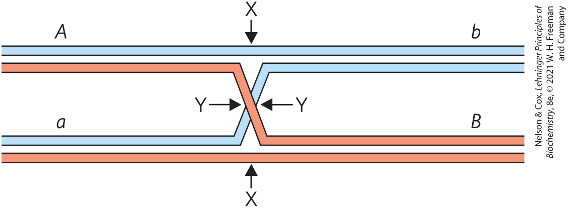
20. Cleavage of Holliday Intermediates A Holliday intermediate forms between two homologous chromosomes, at a point between genes A and B, as shown. The chromosomes have different alleles of the two genes (A and a, B and b). Where would the Holliday intermediate have to be cleaved (points X and/or Y) to generate a chromosome that would carry (a) an Ab genotype or (b) an ab genotype?
21. A Connection between Replication and Site-Specific Recombination Most wild strains of S. cerevisiae have multiple copies of the circular DNA plasmid (named for its contour length of about ), which has ~6,300 bp. For its replication, the plasmid uses the host replication system, under the same strict control as the host cell chromosomes, replicating only once per cell cycle. Replication of the plasmid is bidirectional, with both replication forks initiating at a single, well-defined origin. However, one replication cycle of a plasmid can result in more than two copies of the plasmid, allowing amplification of the plasmid copy number (number of plasmid copies per cell) whenever plasmid segregation at cell division leaves one daughter cell with fewer than the normal complement of plasmid copies. Amplification requires a site-specific recombination system encoded by the plasmid, which serves to invert one part of the plasmid relative to the other. Explain how a site-specific inversion event could result in amplification of the plasmid copy number. (Hint: Consider the situation when replication forks have duplicated one recombination site but not the other.)
 DNA Repair in People with Xeroderma Pigmentosum The condition known as xeroderma pigmentosum (XP) arises from mutations in at least seven different human genes (see
  in
in Data Analysis Problem
22. Mutagenesis in Escherichia coli Many mutagenic compounds act by alkylating the bases in DNA. The alkylating agent R7000 (7-methoxy-2-nitronaphtho[2,1-b]furan) is an extremely potent mutagen.
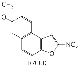
In vivo, R7000 is activated by the enzyme nitroreductase, and this more reactive form covalently attaches to DNA — primarily, but not exclusively, to base pairs.
In a 1996 study, Quillardet, Touati, and Hofnung explored the mechanisms by which R7000 causes mutations in E. coli. They compared the genotoxic activity of R7000 in two strains of E. coli: the wild-type and mutants lacking uvrA activity . They first measured rates of mutagenesis. Rifampicin is an inhibitor of RNA polymerase. In its presence, cells will not grow unless certain mutations occur in the gene encoding RNA polymerase; the appearance of rifampicin-resistant colonies thus provides a useful measure of mutagenesis rates.
The investigators determined the effects of different concentrations of R7000. Their results are shown in the following graph:
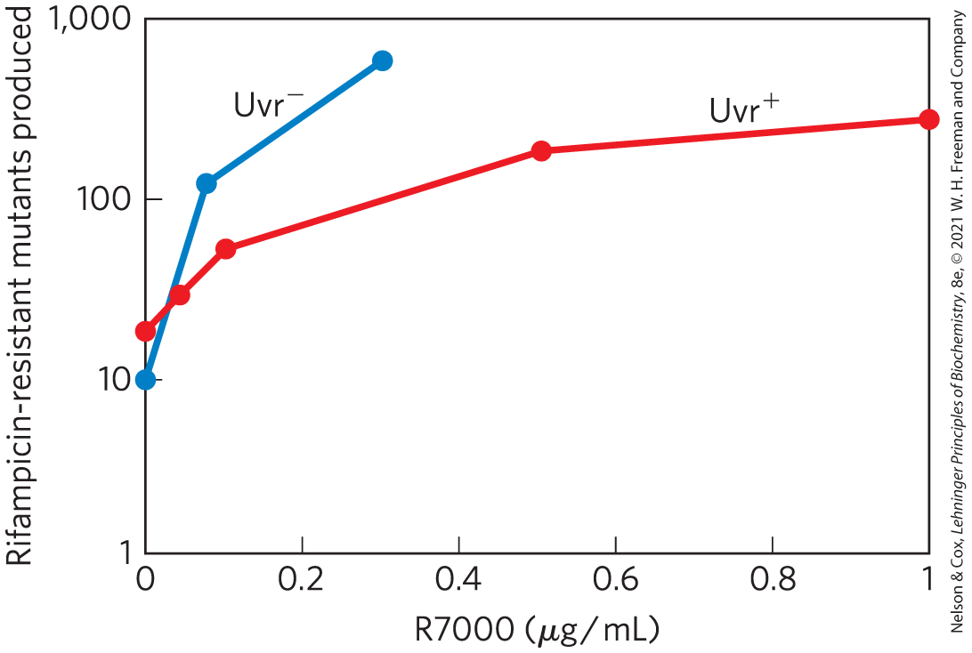
- Why are some mutants produced even when no R7000 is present?
Quillardet and colleagues also measured the survival rate of bacteria treated with different concentrations of R7000, with the following results:
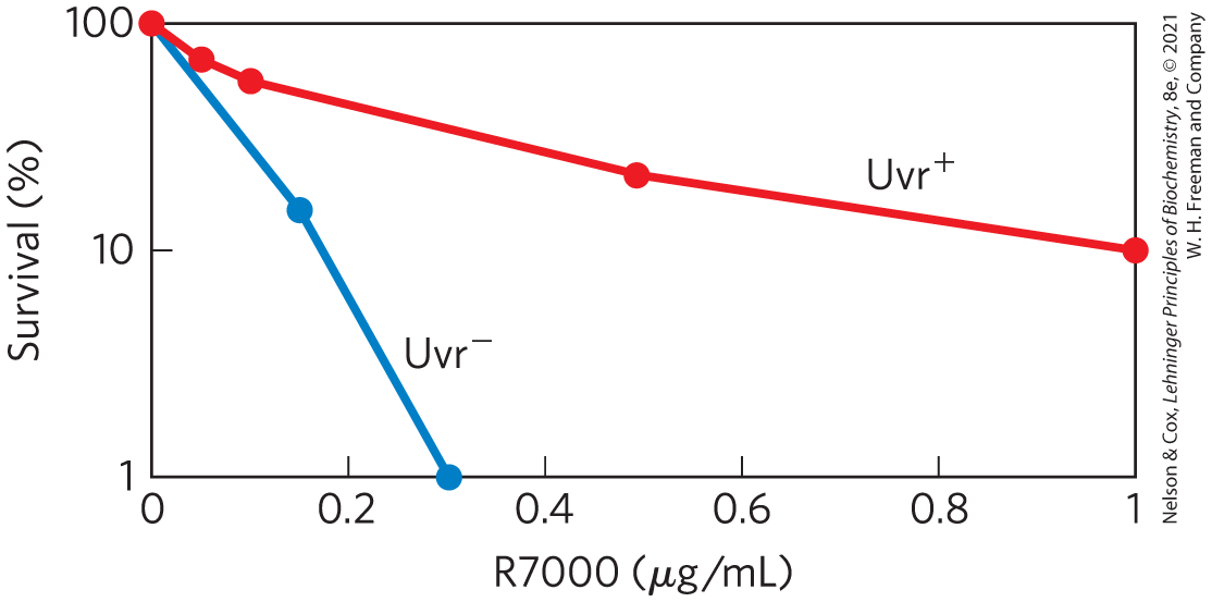
- Explain how treatment with R7000 is lethal to cells.
- Explain the differences in the mutagenesis curves and in the survival curves for the two types of bacteria, and , as shown in the graphs.
The researchers went on to measure the amount of R7000 covalently attached to the DNA in and E. coli. They incubated bacteria with for 10 or 70 minutes, extracted the DNA, and measured its content in counts per minute (cpm) per microgram of DNA.
in DNA
Time (min)
10
76
159
70
69
228
- Explain why the amount of drops over time in the strain and rises over time in the strain.
Quillardet and colleagues then examined the particular DNA sequence changes caused by R7000 in the and bacteria. For this, they used six different strains of E. coli, each with a different point mutation in the lacZ gene, which encodes β-galactosidase. Cells with any of these mutations have a nonfunctional β-galactosidase and are unable to metabolize lactose (i.e., a ). Each type of point mutation required a specific reverse mutation to restore lacZ gene function and . By plating cells on a medium containing lactose as the sole carbon source, the researchers selected for these reverse-mutated, . And by counting the number of following mutagenesis of a particular strain, they could measure the frequency of each type of mutation.
First, they looked at the mutation spectrum in . The following table shows the results for the six strains, CC101 through CC106 (with the point mutation required to produce indicated in parentheses).
CC101 ( to ) CC102 ( to ) CC103 ( to ) CC104 ( to ) CC105 ( to ) CC106 ( to ) 0
0.075
0.15
- Which types of mutation show significant increases above the background rate due to treatment with R7000? Provide a plausible explanation for why some have higher frequencies than others.
- Can all of the mutations you listed in (e) be explained as resulting from covalent attachment of R7000 to a base pair? Explain your reasoning.
- Figure 25-26b shows how methylation of guanine residues can lead to a to mutation. Using a similar pathway, show how an R7000–G adduct could lead to the to or mutations shown above. Which base pairs with the R7000–G adduct?
The results for the bacteria are shown in the following table.
() CC101 ( to ) CC102 ( to ) CC103 ( to ) CC104 ( to ) CC105 ( to ) CC106 ( to ) 0
1
5
- Do these results show that all mutation types are repaired with equal fidelity? Provide a plausible explanation for your answer.
- Why are some mutants produced even when no R7000 is present?
References
- Quillardet, P., E. Touati, and M. Hofnung. 1996. Influence of the uvr-dependent nucleotide-excision repair on DNA adducts formation and mutagenic spectrum of a potent genotoxic agent: 7-methoxy-2-nitronaphtho[2,1-b]furan (R7000). Mutat. Res. 358:113–122.