Chapter Review
KEY TERMS
Terms in bold are defined in the glossary.
- fatty acid
- polyunsaturated fatty acid (PUFA)
- omega-3 (ω-3) fatty acids
- triacylglycerol
- lipases
- phospholipid
- sterols
- glycerophospholipid
- ether lipid
- plasmalogen
- glycolipid
- galactolipid
- sphingolipid
- ceramide
- sphingomyelin
- glycosphingolipid
- cerebroside
- globoside
- ganglioside
- sterol
- cholesterol
- bile acids
- eicosanoid
- prostaglandin (PG)
- thromboxane (TX)
- leukotriene (LT)
- lipoxin (LX)
- vitamin
- vitamin
- cholecalciferol
- vitamin (all-trans-retinol)
- carotenoids
- vitamin E
- tocopherol
- vitamin K
- dolichol
- polyketide
- lipidome
PROBLEMS
1. Operational Definition of Lipids How is the definition of “lipid” different from the types of definitions used for other biomolecules such as amino acids, nucleic acids, and proteins?
2. Structure of an Omega-3 Fatty Acid The omega-3 fatty acid docosahexaenoic acid (DHA, 22:6()) is the most abundant omega-3 fatty acid in the brain and an important component of breast milk. Draw the structure of this fatty acid.
3. Melting Points of Lipids The melting points of a series of 18-carbon fatty acids are stearic acid, ; oleic acid, ; linoleic acid, ; and linolenic acid, .
- What structural aspect of these 18-carbon fatty acids can be correlated with the melting point?
- Draw all the possible triacylglycerols that can be constructed from glycerol, palmitic acid, and oleic acid. Rank them in order of increasing melting point.
- Branched-chain fatty acids are found in some bacterial membrane lipids. Would their presence increase or decrease the fluidity of the membrane (that is, give the lipids a lower or a higher melting point)? Why?
4. Catalytic Hydrogenation of Vegetable Oils Catalytic hydrogenation, used in the food industry, converts double bonds in the fatty acids of the oil triacylglycerols to . How does this affect the physical properties of the oils?
5. Impermeability of Waxes What property of the waxy cuticles that cover plant leaves makes the cuticles impermeable to water?
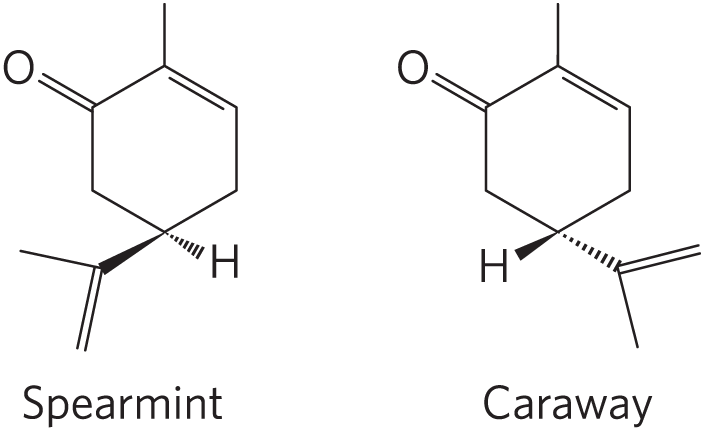
6. Naming Lipid Stereoisomers Carvone, a member of the terpenoid family of chemicals, forms two enantiomers with quite different properties. One enantiomer, abundant in spearmint, smells sweet and minty. The other enantiomer, abundant in caraway seeds, smells spicy and of rye bread. Name the compounds abundant in spearmint and caraway seeds using the RS system.
7. Chemical Reactivity of Lipids Soaps are salts of fatty acids and can be made by mixing triacylglycerols with a strong base such as NaOH. This saponification reaction produces glycerol and fatty acid salts. In a lab experiment, students saponify the triacylglycerol tristearin in the presence of -labeled water. What saponification reaction products will contain the label?
8. Hydrophobic and Hydrophilic Components of Membrane Lipids A common structural feature of membrane lipids is their amphipathic nature. For example, in phosphatidylcholine, the two fatty acid chains are hydrophobic and the phosphocholine head group is hydrophilic. Name the components that serve as the hydrophobic and hydrophilic units for each membrane lipid:
- phosphatidylethanolamine
- sphingomyelin
- galactosylcerebroside
- ganglioside
- cholesterol.
9. Deducing Lipid Structure from Composition A biochemist completely digests a glycerophospholipid with a mixture of phospholipases A and D. HPLC and MS analysis reveals the presence of an amino acid of 105.09 Da, a saturated fatty acid of 256.43 Da, and an omega-3 monounsaturated fatty acid of 282.45 Da. Which amino acid does the glycerophospholipid contain? Draw the most likely structure of this glycerophospholipid.
10. Deducing Lipid Structure from Molar Ratio of Components Complete hydrolysis of a glycerophospholipid yields glycerol, two fatty acids (16:1() and 16:0), phosphoric acid, and serine in the molar ratio 1:1:1:1:1. Name this lipid and draw its structure.
11. Lipids in Blood Group Determination We note in Figure 10-13 that the structure of glycosphingolipids determines the blood groups A, B, and O in humans. It is also true that glycoproteins determine blood groups. How can both statements be true?
12. The Action of Phospholipases The venom of the Eastern diamondback rattler and the Indian cobra contains phospholipase , which catalyzes the hydrolysis of fatty acids at the C-2 position of glycerophospholipids. The phospholipid breakdown product of this reaction is lysolecithin, which is derived from phosphatidylcholine. At high concentrations, this and other lysophospholipids act as detergents, dissolving the membranes of erythrocytes and lysing the cells. Extensive hemolysis may be life-threatening.
- All detergents are amphipathic. What are the hydrophilic and hydrophobic portions of lysolecithin?
- The pain and inflammation caused by a snake bite can be treated with certain steroids. What is the basis of this treatment?
- Though the high levels of phospholipase in venom can be deadly, this enzyme is necessary for a variety of normal metabolic processes. What are these processes?
13. Intracellular Messengers from Phosphatidylinositols The hormone vasopressin is an extracellular signal that activates a specific phospholipase C in the membrane. Cleavage of by phospholipase C generates two products. What are they? Compare their properties and their solubilities in water, and predict whether either would diffuse readily through the cytosol.
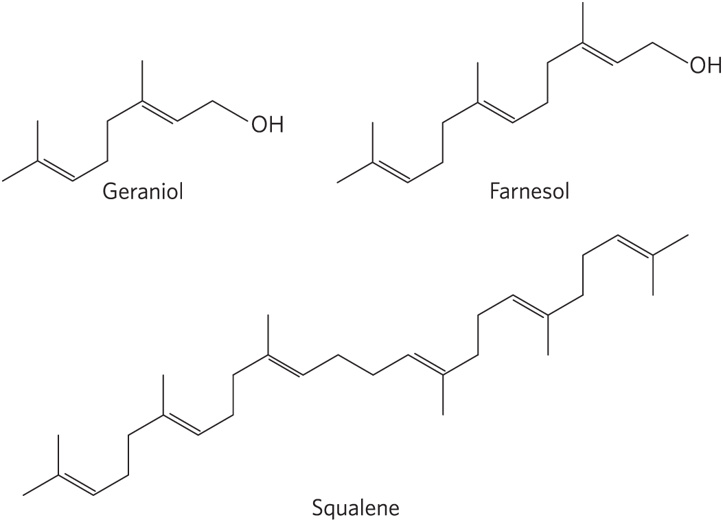
14. Isoprene Units in Isoprenoids Geraniol, farnesol, and squalene are called isoprenoids because they are synthesized from five-carbon isoprene units. In each compound, circle the five-carbon units representing isoprene units (see Fig. 10-22).
15. Hydrolysis of Lipids Name the products of mild hydrolysis with dilute NaOH of
- 1-stearoyl-2,3-dipalmitoylglycerol
- 1-palmitoyl-2-oleoylphosphatidylcholine.
16. Effect of Polarity on Solubility Rank a triacylglycerol, a diacylglycerol, and a monoacylglycerol in order of decreasing solubility in water. Assume that each acylglycerol contains only palmitic acid.
17. Chromatographic Separation of Lipids Suppose that you apply a mixture of lipids to a silica gel column and then wash the column with increasingly polar solvents. The mixture consists of phosphatidylserine, phosphatidylethanolamine, phosphatidylcholine, cholesteryl palmitate (a sterol ester), sphingomyelin, palmitate, n-tetradecanol, triacylglycerol, and cholesterol. In what order will the lipids elute from the column? Explain your reasoning.
18. Identification of Unknown Lipids Johann Thudichum, who practiced medicine in London about 100 years ago, also dabbled in lipid chemistry in his spare time. He isolated a variety of lipids from neural tissue and characterized and named many of them. His carefully sealed and labeled vials of isolated lipids were rediscovered many years later.
- How would you confirm, using techniques not available to Thudichum, that the vials labeled “sphingomyelin” and “cerebroside” actually contain these compounds?
- How would you distinguish sphingomyelin from phosphatidylcholine by chemical, physical, or enzymatic tests?

BIOCHEMISTRY ONLINE
19. Using the LIPID MAPS Database to Find Solubility Information Lipidomics has identified thousands of cellular lipids. LIPID MAPS is an online database containing over 40,000 unique lipid structures, as well as information on the chemical and physical properties of each lipid (www.lipidmaps.org). One important parameter when working with lipids is log P, where P is the octanol:water partition coefficient, an indicator of lipophilicity.
- Look up cholesterol, sphingosine, linoleic acid, and stearic acid in LIPID MAPS and use the reported log P values to place them in order of increasing solubility in octanol.
- Pharmacologists often study log P values when developing new drugs. Why would knowing a drug’s log P value be informative?
20. Characteristics of Lipid Transport Proteins Often when lipids are transported between different tissues, they are carried by proteins. In this exercise, you will explore the interactions between a lipid and a protein using the PDB (www.rcsb.org). Use the PDB identifier 2YG2 and study the structure of the complex between HDL-associated apolipoprotein M and sphingosine-1-phosphate. Navigate to 3D View: Structure to answer the following questions.
- What protein motif is adopted by apolipoprotein M?
- Which amino acid residues do you find lining the sphingosine binding pocket? What do they have in common?
- The phosphoryl group of sphingosine-1-phosphate is exposed on the surface of the protein. Why do you suppose it is important that the transport protein binds the hydrocarbon tail of sphingosine-1-phosphate but not necessarily the polar head group?
DATA ANALYSIS PROBLEM
21. Determining the Structure of the Abnormal Lipid in Tay-Sachs Disease Box 10-1, Figure 1, shows the pathway of breakdown of gangliosides in healthy (normal) individuals and in individuals with certain genetic diseases. Some of the data on which the figure is based were presented in a paper by Lars Svennerholm (1962). Note that the sugar Neu5Ac, N-acetylneuraminic acid, represented in the Box 10-1 figure as , is a sialic acid.
Svennerholm reported that “about 90% of the monosialogangliosides isolated from normal human brain” consisted of a compound with ceramide, hexose, N-acetylgalactosamine, and N-acetylneuraminic acid in the molar ratio 1:3:1:1.
- Which of the gangliosides (GM1 through GM3 and globoside) in Box 10-1, Figure 1, fits this description? Explain your reasoning.
- Svennerholm reported that 90% of the gangliosides from a patient with Tay-Sachs disease had a molar ratio (of the same four components given above) of 1:2:1:1. Is this consistent with the Box 10-1 figure? Explain your reasoning.
To determine the structure in more detail, Svennerholm treated the gangliosides with neuraminidase to remove the N-acetylneuraminic acid. This resulted in an asialoganglioside that was much easier to analyze. He hydrolyzed it with acid, collected the ceramide-containing products, and determined the molar ratio of the sugars in each product. He did this for both the normal gangliosides and the Tay-Sachs gangliosides. His results are shown below.
Ganglioside Ceramide Glucose Galactose Galactosamine Normal
Fragment 1
1
1
0
0
Fragment 2
1
1
1
0
Fragment 3
1
1
1
1
Fragment 4
1
1
2
1
Tay-Sachs
Fragment 1
1
1
0
0
Fragment 2
1
1
1
0
Fragment 3
1
1
1
1
- Based on these data, what can you conclude about the structure of the normal ganglioside? Is this consistent with the structure in Box 10-1? Explain your reasoning.
- What can you conclude about the structure of the Tay-Sachs ganglioside? Is this consistent with the structure in Box 10-1? Explain your reasoning.
Svennerholm also reported the work of other researchers who “permethylated” the normal asialoganglioside. Permethylation is the same as exhaustive methylation: a methyl group is added to every free hydroxyl group on a sugar. They found the following permethylated sugars: 2,3,6-trimethylglycopyranose; 2,3,4,6-tetramethylgalactopyranose; 2,4,6-trimethylgalactopyranose; and 4,6-dimethyl-2-deoxy-2-aminogalactopyranose.
- To which sugar of GM1 does each of the permethylated sugars correspond? Explain your reasoning.
- Based on all the data presented so far, what pieces of information about normal ganglioside structure are missing?
 , is a sialic acid.
Reference
- Svennerholm, L. 1962. The chemical structure of normal human brain and Tay-Sachs gangliosides. Biochem. Biophys. Res. Comm. 9:436–441.