the heart does calcium-induced-calcium-release , skeletal muscle uses direct contact of ryanodine receptors instead
How to Increase Conduction Velocity :
add myelin
increases resistance ( less leak )
decreases capacitance ( more distance between inside and outside of cell )
increase length between internodes
fewer nodes = fewer sites where the AP needs to regenerate , saving time
increase internal diameter ( giant squid )
lower cytosolic resistance ,
allows charges to diffuse easier / faster
Acute Inflammatory Demyelinating Polyradiculoneuropathy ( AIDP )
autoimmune attack demyelination on PNS nerves
Guillain-Barre type
Multiple Sclerosis ( MS ) :
autoimmune attack on oligodendrocytes ( CNS )
loss of myelin
disrupted paranodal and nodal organization
treatment = K+ channel blocker
Myasthenia Gravis ( MG ) :
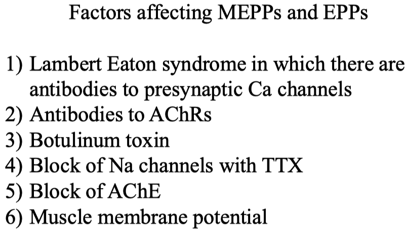
autoimmune attack on skeletal muscle neuromuscular junction ACh receptors
treatment = acytlecholine esterase inhibitors
but the balance is tricky , because too much inhibitor will cause too much available acetylcholine
then the receptors will naturally become desensitized on their own due to excess ACh
If you voltage clamp some muscle fiber at -90 mV vs clamping it at -20 mV ,
the one clamped at -90 will have a much larger current measured
there is a larger driving force for acetylcholine
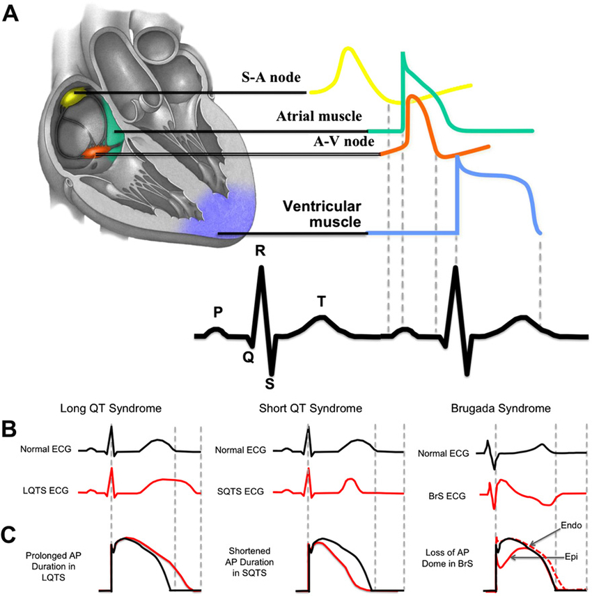
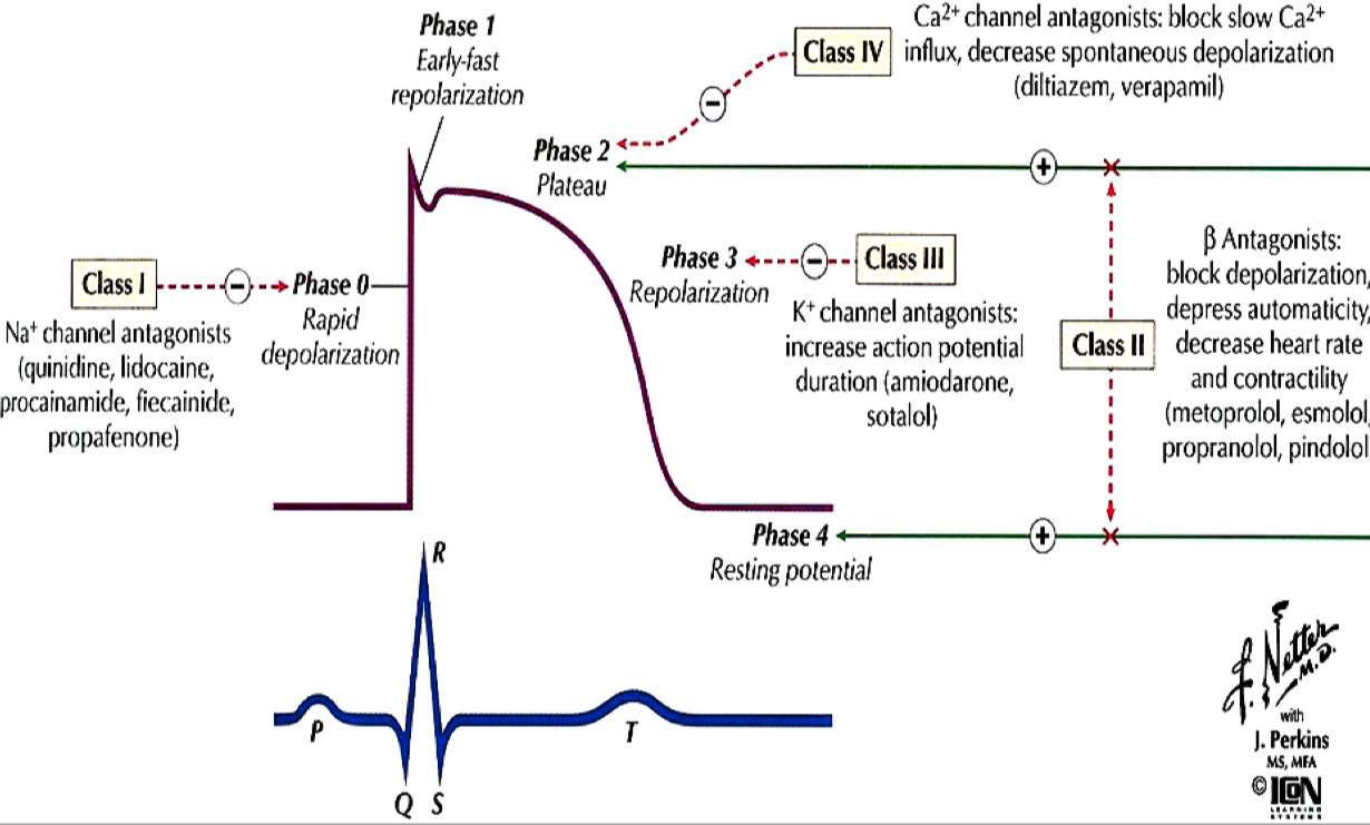
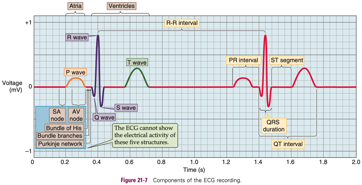
| Neurotransmitter | Receptor Type | Subtype Name | Other Names / Notes | Ions Conducted | Effect |
|---|---|---|---|---|---|
| Acetylcholine (ACh) | Ionotropic | Nicotinic | Ligand-gated ion channel | Na+ in , K+ out ( some also Ca2+ permeable ) | Excitatory |
| Metabotropic | Muscarinic | GPCR ( M1–M5 subtypes ) | — ( via second messengers ) | Usually excitatory ( M1 , M3 , M5 ) or inhibitory ( M2 , M4 ) depending on subtype | |
| Glutamate | Ionotropic | AMPA , Kainate | Ligand-gated ion channels | Na+ in, K+ out | Excitatory |
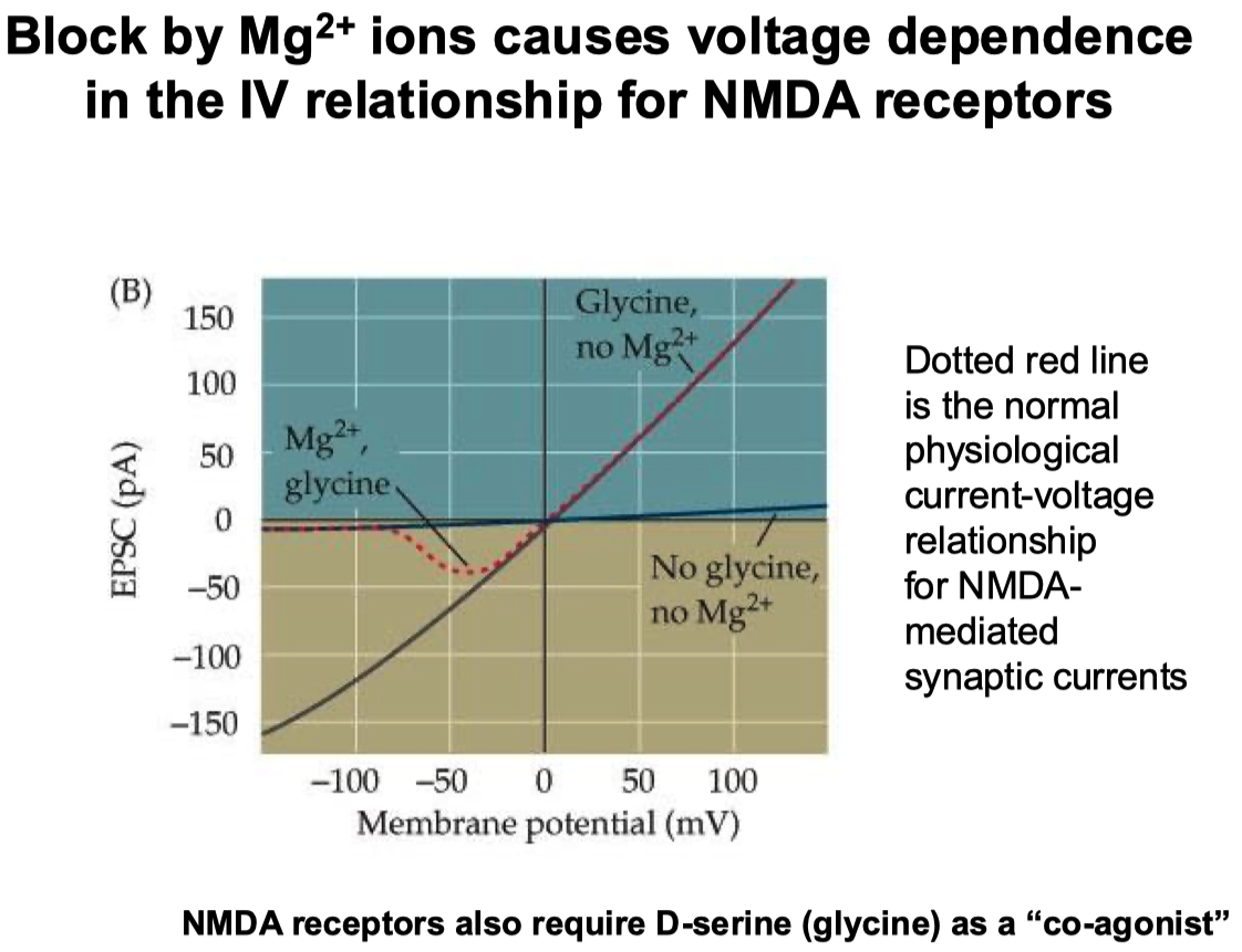
| Ionotropic | NMDA | Requires glycine co-agonist , voltage-gated Mg2+ block | Na+, Ca2+ in, K+ out | Strong excitatory , Ca2+-dependent signaling | |
| Metabotropic | mGluR ( types 1–8 ) | GPCRs | — ( via second messengers ) | Modulatory ( can be excitatory or inhibitory ) | |
| GABA | Ionotropic | GABAA | Ligand-gated Cl– channel | Cl– in ( hyperpolarizes ) | Inhibitory |
| Metabotropic | GABAB | GPCR activating K+ channels, inhibiting Ca2+ channels | ↑ K+ out , ↓ Ca2+ in ( via second messenger ) | Inhibitory | |
| Glycine | Ionotropic | Glycine receptor | Ligand-gated Cl– channel | Cl– in ( hyperpolarizes ) | Inhibitory |
| Metabotropic | — ( none ) | No known metabotropic receptor | — | — |
| Receptor | Kinetics | Calcium Permeable ? | Voltage Dependent ? |
|---|---|---|---|
| AMPA | Fast | No | No |
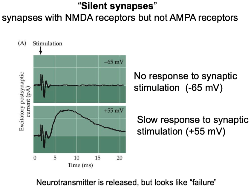
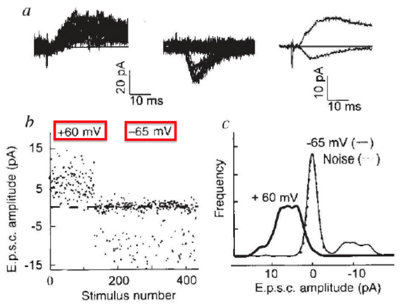
| NMDA | Slow | Yes | Yes ( via magnesium block ) |
Dopamine for example can bind to both stimulatory and inhibitory GPCRs
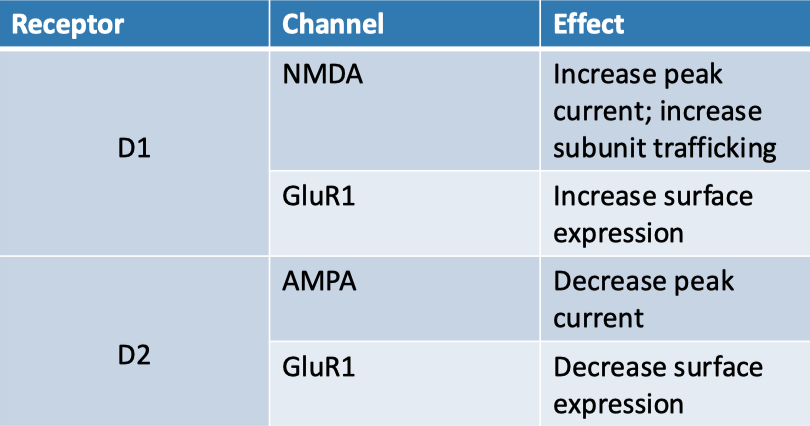
Extracellular magnesium is used to block / inactivate NMDA channels at negative potentials
Heuser Paper :
Hypothesis = synaptic vesicle exocytosis underlies quantal neurotransmitter release
one synaptic vesicle fusing with the membrane corresponds to one quantum of neurotransmitter release,
and these events occur independently at the presynaptic active zone
Conclusions :
Each vesicle undergoing exocytosis corresponds to a quantum of neurotransmitter release
Vesicle fusion events are independent, not cooperative
Quick-freezing successfully captures exocytosis events.
6At low levels of release, collapsed vesicles must be included to match quantal output
Evidence :
Freeze-fracture electron microscopy captured vesicles fusing with the membrane 5–6 ms after nerve stimulation, matching the timing of neurotransmitter release.
4-aminopyridine (4-AP) was used to increase quantal output, showing a dose-dependent increase in vesicle exocytosis events.
Quantitative correlation: The number of vesicles fusing matched the number of quanta released, across low to high 4-AP concentrations.
Spatial analysis showed vesicle openings occur precisely at active zones, not elsewhere, and are randomly distributed, supporting independent vesicle fusion.
At lower 4-AP doses, inclusion of collapsed vesicles (early fusion stages) improved correlation with physiological data.
No evidence for synchronized or cooperative fusion — each vesicle acts independently.
Together, this evidence strongly supports the hypothesis that synaptic vesicles fuse with the membrane to release neurotransmitter quanta, and that this can be directly visualized and quantified using high-resolution, time-locked quick-freezing techniques.
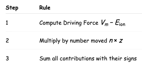
How to calculate
| Feature | Synaptobrevin ( zipper ) KO (VAMP KO) | Synaptotagmin ( sensor / brake ) KO |
|---|---|---|
| Evoked Amplitude | ↓↓↓ ( almost abolished ) | ↓↓↓ ( almost abolished ) |
| Evoked Frequency | ↓↓↓ | ↓↓↓ |
| Mini Amplitude | ↓ ( reduced ) | increased |
| Mini Frequency | ↓↓↓ ( very rare ) | ↑ ( increased spontaneous fusion ) |
| Docking | unaffected , they still dock | decreased |
| Fusion | they don't fuse | Ca²⁺-triggered fusion defective; spontaneous fusion increased |
| Protein/Channel | Location | Direction (From → To) | What is Moved |
|---|---|---|---|
| ORAI1 | Plasma membrane | Extracellular space → Cytosol | Ca²⁺ (influx) |
| STIM1 | ER membrane | — | Senses low ER Ca²⁺ |
| TRPC | Plasma membrane | Extracellular space → Cytosol | Ca²⁺ / Na⁺ (non-selective cation) |
| PMCA | Plasma membrane | Cytosol → Extracellular space | Ca²⁺ (ATP-dependent) |
| SERCA | ER membrane | Cytosol → ER lumen | Ca²⁺ (ATP-dependent) |
| RyR | ER membrane | ER lumen → Cytosol | Ca²⁺ (CICR) |
| Ins(1,4,5)P₃R | ER membrane | ER lumen → Cytosol | Ca²⁺ (via IP₃) |
| MCU complex | Mitochondrial inner membrane | Cytosol → Mitochondrial matrix | Ca²⁺ |
| VDAC | Mitochondrial outer membrane | Cytosol → Intermembrane space | Ca²⁺ and other metabolites |
| TRPML1 | Lysosomal membrane | Lysosome → Cytosol | Ca²⁺ |
| Cav channel | Plasma membrane | Extracellular space → Cytosol | Ca²⁺ (voltage-gated) |
| BKCa channel | Plasma membrane | Cytosol → Extracellular space | K⁺ (Ca²⁺-activated) |
| CaCC channel | Plasma membrane | Cytosol → Extracellular space | Cl⁻ (Ca²⁺-activated) |
| IP₃R | SR membrane | SR lumen → Cytosol | Ca²⁺ (via IP₃) |
| EAAT1/2 | Astrocyte membrane | Synaptic cleft → Astrocyte cytosol | Glutamate + 2 Na⁺ + H⁺ in, K⁺ out |
| EAAT3 | Neuron membrane | Synaptic cleft → Neuron cytosol | Glutamate + 2 Na⁺ + H⁺ in, K⁺ out |
| SNAT1/2 | Neuron membrane | Extracellular → Neuron cytosol | Glutamine + Na⁺ |
| SNAT3/5 | Astrocyte membrane | Bidirectional (mostly cytosol → out) | Glutamine + Na⁺ / H⁺ exchanger |
| Na⁺/K⁺ ATPase | Neuron membrane | 3 Na⁺ out / 2 K⁺ in (cyclic) | Maintains gradient (ATP-dependent) |