Links
Field of View
Resolution and Numerical Aperture
Chromatic Aberration
Working Distance
Intro
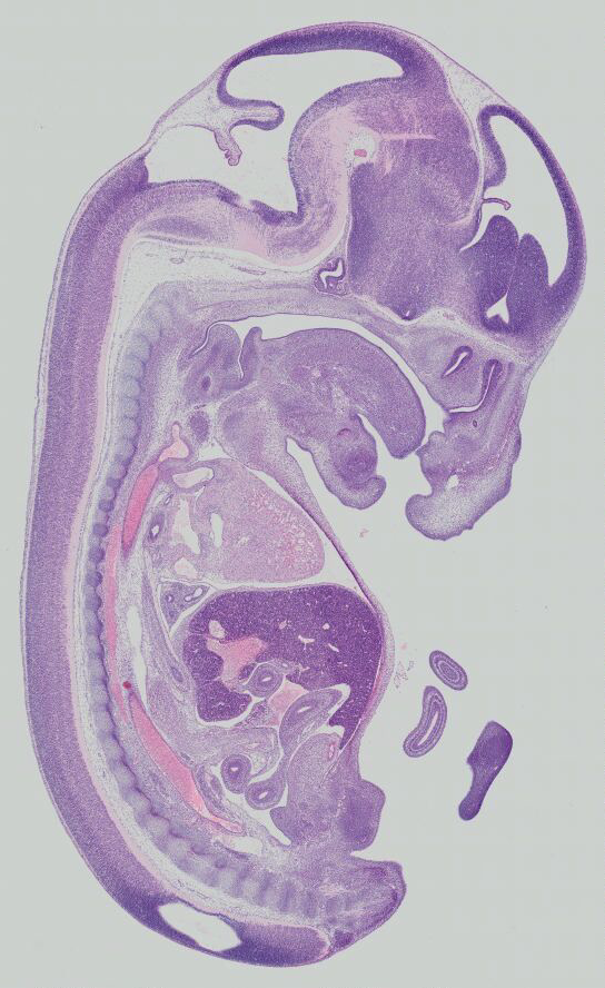
Your first assignment as a research assistant in a new lab is to image and document sections of a mouse embryo.
Because the embryo is small, the animal has been sectioned intact along the sagittal plane.
Other workers in the lab have already prepared the samples for you.
Each section of the embryo is 250 μm thick.
These sections were then stained, mounted on standard microscope slides, and cover slipped.
Before beginning this task, you will need to be aware of the strengths and limitations of the microscopy equipment you have on hand.
With this information you can then make appropriate decisions on how to image these samples to efficiently obtain the relevant data from these slides.
You should refer to this online viewer to assist you in working through these decisions:
Let Image Width =
Let Image Height =
Notes
Objective = series of lenses that keeps image flat and colors constant
You are always limited by what you can see by field number of eye piece
- eye piece always has the smallest field number compared to objective lens
- Rectangular viewports have independent fields of view for length and width.
- Whereas for the eyepiece , we can just use the area formula for a circle
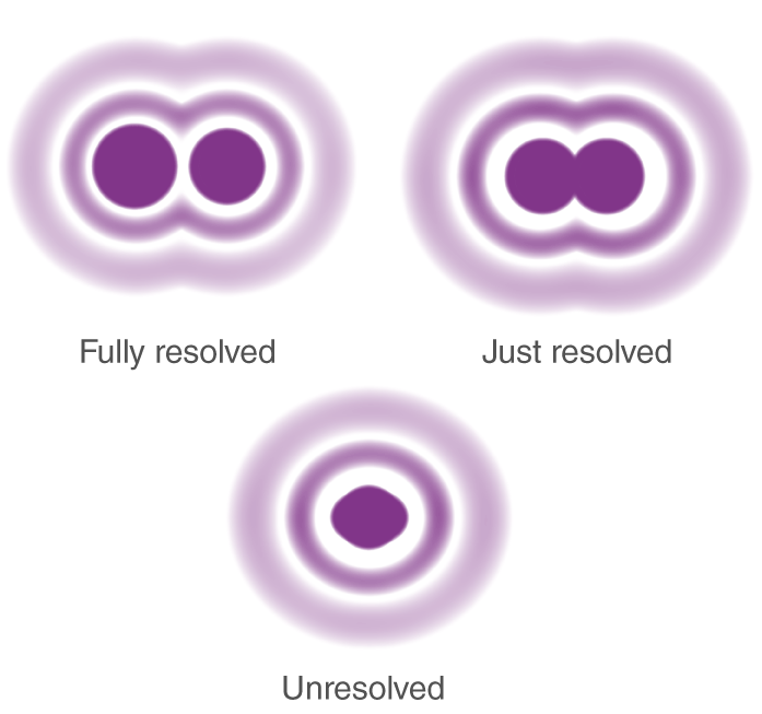
Resolution = "distance between 2 points where you can still tell there are 2 objects"
"you can buy a very low resolution optic , with a really high
"we need to see at least 3 microns or 3000 nano meters"
- "so we need a resolution of < 3000 nm"
Topic 1: Field of View
How big this embryo section?
- (zoom out; right click on image to access measurement mode; pick two points)
Come up with answer in mm2
How much of the embryo will be visible through the microscope at a time?
- What is the field number ( FN ) of the different objectives available on the microscope?
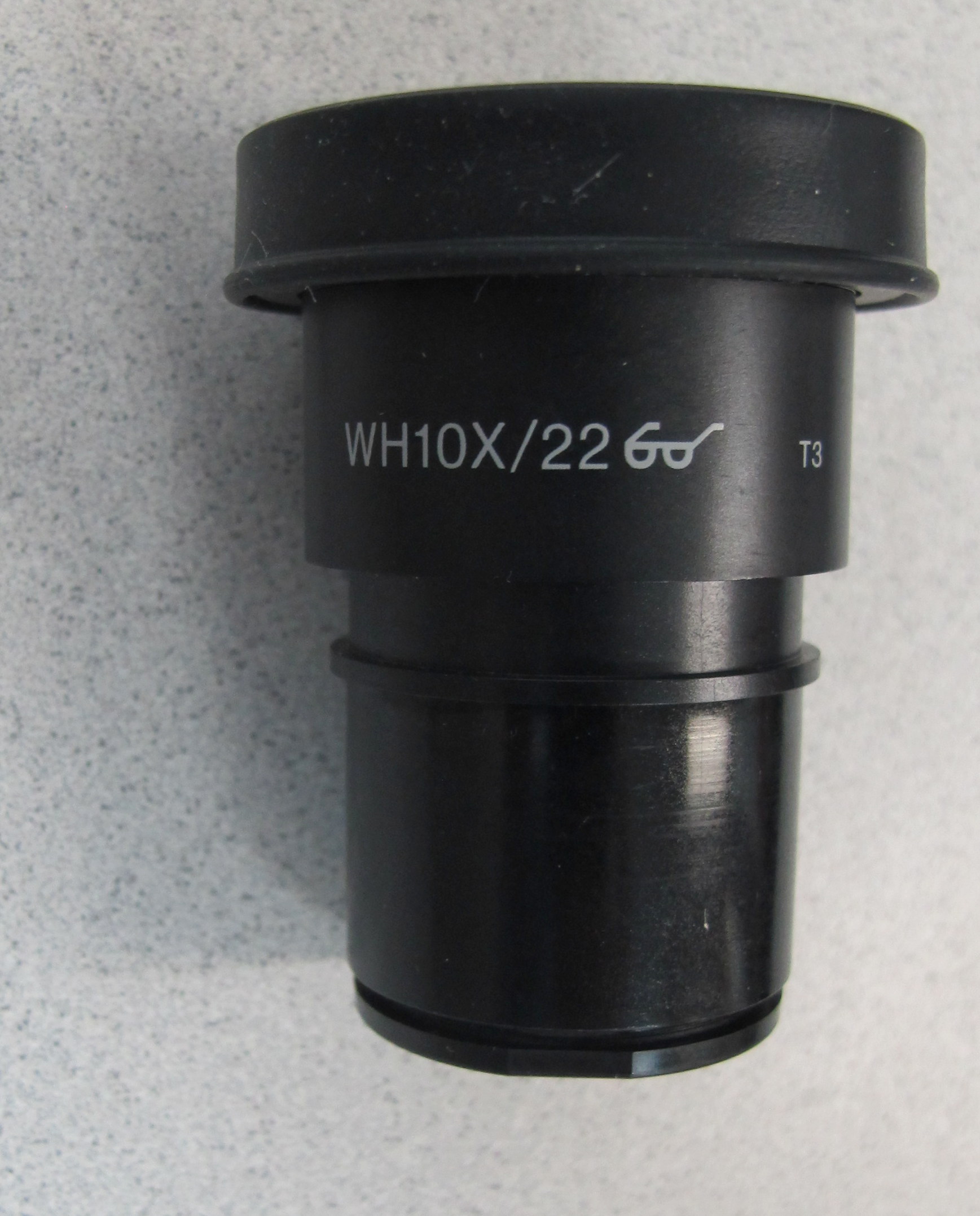
This is an image of the eye piece on your microscope.
- Magnification =
- Field Number =
- Magnification =
If the objective and eye piece field numbers do not match ,
- your image will be limited by the smaller of the two numbers
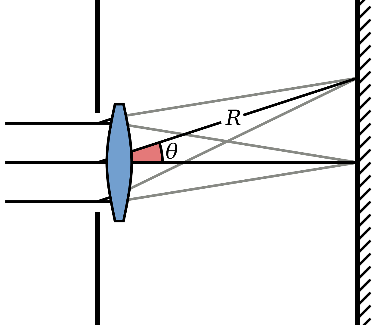
Calculate the field of view ( in mm ) for the following objectives :
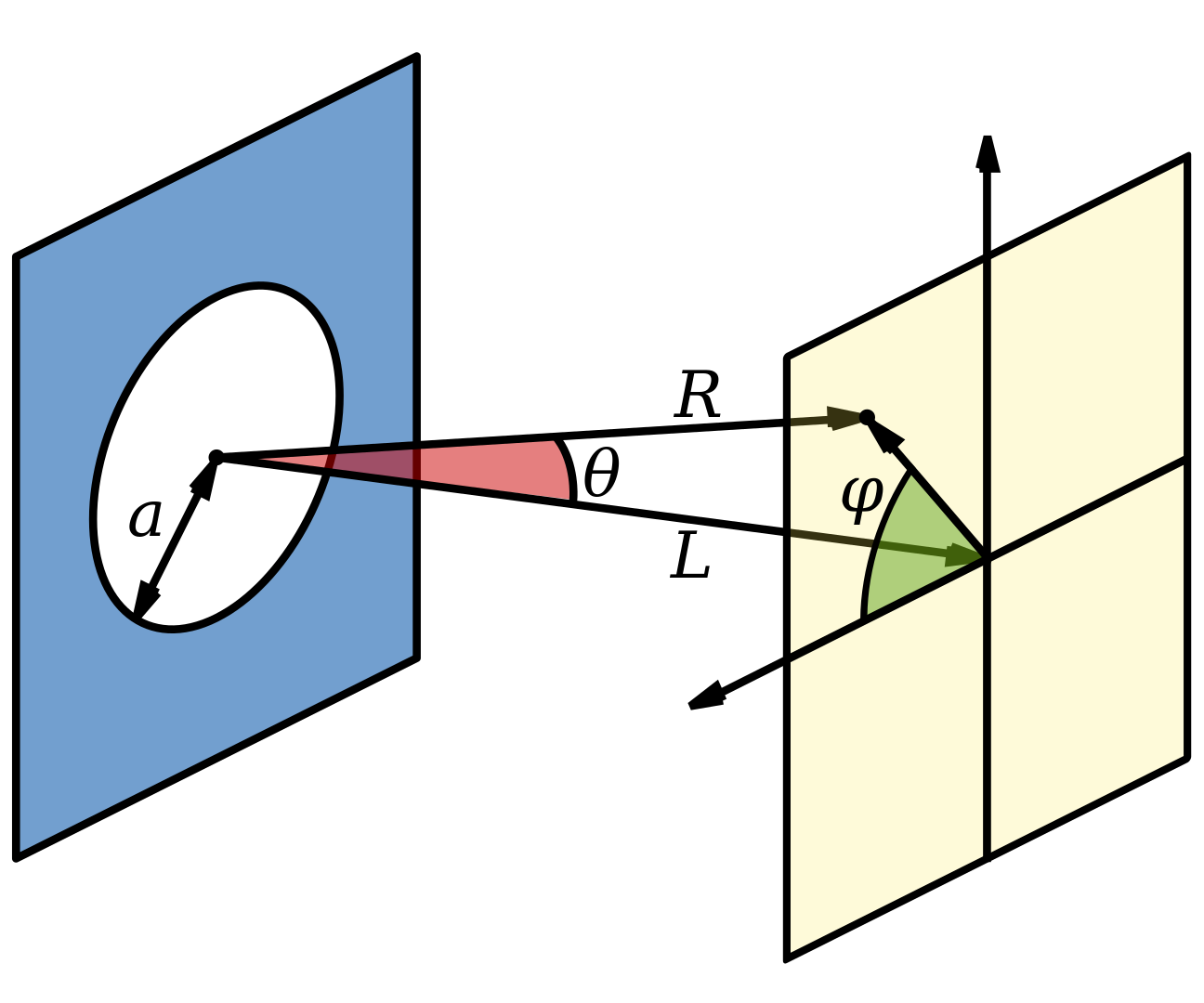
The FOV represents the diameter of the circle visible through the eyepieces.
Calculate the visible area ( in mm2 ) and percent of total area of the embryo it represents for the following objectives :
The digital cameras attached to microscopes have square or rectangular sensors
- so they won’t capture everything you can see by eye through the microscope.
Your lab microscope is equipped with a SPOT RT3 camera.
Using the specification sheet, determine the effective field number ( FN ) of this camera.

What percent of the embryo will be captured in each image you would take using a 10x and 60x objective?
How many images will be necessary to document the whole embryo?
Topic 2: Resolution and Numerical Aperture
Rayleigh Equations :
- https://en.wikipedia.org/wiki/Airy_disk#Mathematical_formulation
- https://mathworld.wolfram.com/BesselFunctionoftheFirstKind.html
- https://physics.stackexchange.com/questions/264925/where-does-the-0-61-come-from-in-r-sin-theta-0-61-lambda
- https://byjus.com/physics/rayleigh-criterion/
- http://kmdouglass.github.io/posts/simple-pupil-function-calculations/
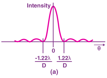
Where :
- ie , the angle between the axis of the circular aperture and the line between aperture center and observation point
Where :
- https://en.wikipedia.org/wiki/Bessel_function#Bessel_functions_of_the_first_kind:_J%CE%B1
- Let alpha (
Where :
Solving for first zero ( Wolfram Alpha ):
Let Wave Number (
- Where
- Where
Assume
Isolating for
- Putting everything back in terms of resolution via the Rayleigh equation :
While collecting images with the 60X objective will require significantly more time , it may be necessary depending on the level of detail you wish to capture.
- This means you’ll need to know something about the resolving power of your microscope.
Zoom in again on the embryo online at emouseatlas.org.
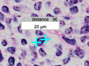
What is the size of some of the smallest objects or features in the image?
What kind of resolution would you need to achieve to replicate this level of detail?
- We need the resolution to be smaller than
Solving for
Solving for
- We need the resolution to be smaller than
Using light at 550nm , calculate the resolution possible using objectives with the following numerical apertures ( NA ).
Review the NA of the objectives available on the lab microscope.
Do your calculations influence or limit your choice of objectives? Why or why not?
UPLFLN 10X2 :
- W.D. = 10.0 , MAG = 10x , F.N. = 26.5 , NA = 0.3 , IM = none
UPLSAPO 10X2 :
- W.D. = 3.1 , MAG = 10x , F.N. = 26.5 , NA = 0.4 , IM = none
UPLSAPO 20X :
- W.D. = 0.6 , MAG = 20x , F.N. = 26.5 , NA = 0.75 , IM = none
UPLSAPO 40X2 :
- W.D. = 0.18 , MAG = 40x , F.N. = 26.5 , NA = 0.95 , IM = none
UPLSAPO 60XO :
- W.D. = 0.15 , MAG = 60x , F.N. = 26.5 , NA = 1.35 , IM = oil ( n = 1.515 )
UPLSAPO 100XS :
- W.D. = 0.2 , MAG = 100x , F.N. = 22.0 , NA = 1.35 , IM = silicon oil ( n = 1.45 to 1.50 ( source ) )
SPOTTM RT3 2.0 MP Slider :
- FOV = 11.8 mm x 8.9 mm
The wavelength of light is a key parameter in the equation for resolution.
This suggests the choice of wavelength influences the resolution possible for your microscope.
Calculate the maximum resolution possible with a high numerical aperture ( 1.40 ) objective.
Using the equation for NA (
- let refractive index of air = 1.00
Explain why.
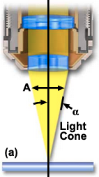
- Max Half-Angle light can enter into objective is
- Max Half-Angle light can enter into objective is
Topic 3: Aberrations
Distance from the lense where everything comes into focus.
Big lens with smooth surface has difference than small lense with large curvature.
Different wavelengths as they pass thorugh the material have different refractive indices.
- Different wavelengths converge at different focal points
Chromatically corrected lens focuses everything at the correct place
Fluorite = cheapest lens
Aberration corrections and numerical drive the price of the lens
What kind of staining is used on this embryo tissue?
Stain = haematoxylin and eosin
Use the simplified lens maker formula to calculate the changes in focal length of a lens for the wavelengths listed
Interactive Lens-Marker's Equation :
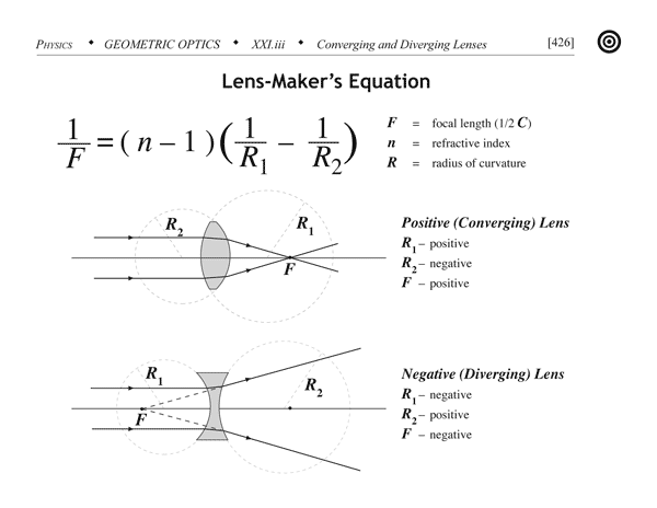
Let
Red light ( n = 1.513 ) :
- Violet light ( n = 1.532 ) :
Will these differences affect your imaging?
- Violet focuses in front of the focal point
- Red focuses further away from the focal point than violet light
- Spherical aberration , anything on the edges gets distorted
Topic 4: Working Distance
- Every objective has a limited working distance.
You must keep this in mind when choosing objectives for your studies.
In order to safely focus on any region of your sample ,
- the sum of the tissue sample thickness along with the coverslip thickness must be less than the working distance of the objective.
Your coworker who prepared the embryo sections informs you that
Working Distance = Coverslip thickness + distance to objective
Coverslip types and thickness tolerances :
- (
- (
Review the spec sheets for the objective on your microscope.
So we need a working distance bigger than 0.42 mm
Are there any limitations on which objectives can be used to image the embryo sections?
UPLSAPO 40X2 , UPLSAPO 60XO , UPLSAPO 100XS have working distances smaller than
- so they will NOT work with our cover slip and tissue thickness
Explain why.
- Only UPLFLN 10X2 , UPLSAPO 10X2 , UPLSAPO 20X lenses have a working distance larger than
- as you get higher magnification , the working distance goes down
- Only UPLFLN 10X2 , UPLSAPO 10X2 , UPLSAPO 20X lenses have a working distance larger than
Review the spec sheets for the objectives available on your microscope.
Is there a relationship between NA and working distance? Explain why or why not.
https://en.wikipedia.org/wiki/Numerical_aperture
- Increasing the magnification and the numerical aperture of the objective reduces the working distance
https://physics.stackexchange.com/a/528093
For a given numerical aperture , one can usually increase the working distance
- but it requires making all the lens elements larger
For a given numerical aperture , there is a tradeoff between working distance and cost
- because larger elements will cost more.
Lastly , summarize your strategy for documenting these embryo sections.
What objective( s ) are best?
- UPLSAPO 10X2
Why did you choose them?
- want to be able to focus through the whole specimen , and take as little images as possible
- UPLSAPO 10X2 has higher NA so resolution is lower ( "better resolution" )
- apo corrected