Differences between using Fluorescence and Brightfield
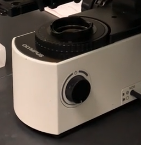
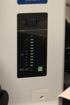
When using fluorescence , you don't have to turn on the light source that is part of the microscope
aka , don't turn this on :
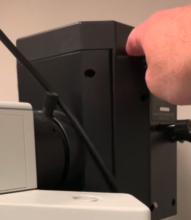
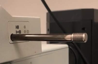
We are doing "epifluoresence" , and the light source comes from the back ( mercury halide lamp ) :
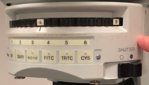
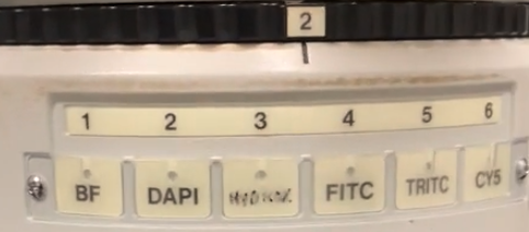
Light path travels into "filter turret" , where you can select different filters to use :
Then light passes down into the objective , where it illuminates the sample :
the fluorescent light is then collected by the objective , and reflected back up into the eye piece or camera
Don't have to mess with preset button ( just leave it turned off ) :
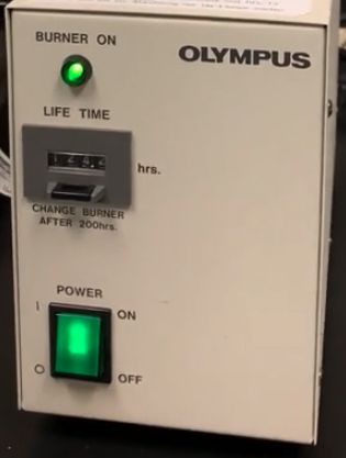
However , the mercury burner lamp still needs to be turned on when you first get to the microscope :
- make sure "burner light" is solid green
Sample we are using is in the back fridge
its a special sample they purchased of "cultured cells"
labeled with 3 different fluorescent markers
- Beta Tubulin
- Flamoydin ?
- DAPI = for nucleu
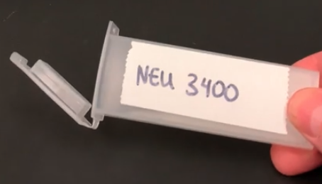
MAKE SURE THE SAMPLE CONTAINER STAYS COLD
WHEN YOU ARE DONE WITH THE SLIDE , PUT IT BACK IN THE TUBE , AND THEN BACK IN THE FRIDGE
- otherwise it will fade
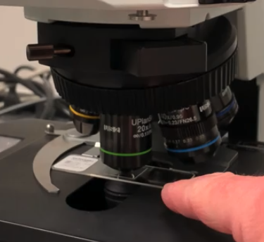
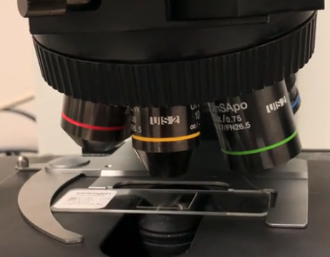
Start with the 10x , and focus and find your sample
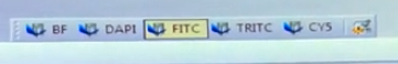
Select appropriate channel / filter
- use FITC ( blue light ) to see green fluorescence
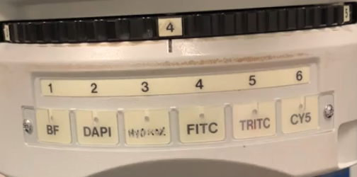
- use TRITC ( green light ) to see red fluorescence
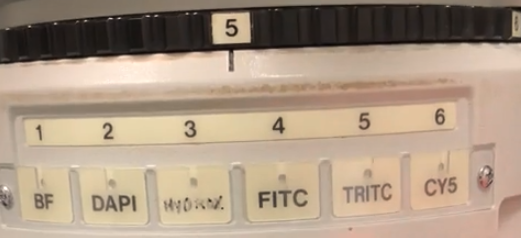
DAPI ( blue light ) to see nuclei
- chemical that labels double stranded DNA
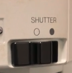
To illuminate the sample , open the shutter
Once you have the sample in good focus , take pictures with 20x and 40x objectives
Pull light selector knob all the way out for fluorescence
- the images will be much dimmer than they were for bright field
When you are in the fluorescence mode and using the CellSens software :
make sure you choose the appropriate acquisition setting :
Adjust exposure time so you can see the sample
if you sit down to the computer chair and you can't see anything on the computer screen
but you CAN see something via the eyepiece
- then your exposure settings are messed up ( probably too low ) , so turn up
Ctrl+Hkeyboard shortcut = toggles "high/low" modeapplies lookup table to pixels
totally dark pixels are turned to blue color
saturated pixels are turned to red color
- so minimize red color via exposure setting
toggle back and forth between "high/low" mode to get reasonable image
Save image file as usual
might have to adjust focus as you change different filters
CLOSE THE SHUTTER once you are done imaging
increases the lifespan of the slide
especially with DAPI ( uses short blue wavelengths )
- can bleach the sample
You can merge all three images ( if you want ) , all of the channels have to be open at the same time

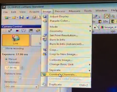
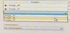
make sure you select "convert to RGB"
click "ok"
- creates a new merged image