Sensitization in Aplysia
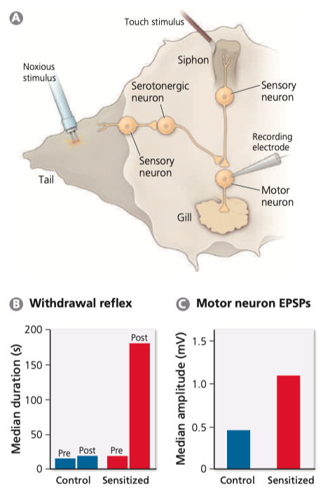
Figure 3.1 - Sensitization of Aplysia's Withdrawal Reflex

- Depicted in (A) is a semi-intact Aplysia preparation
- The bar graph in (B) reveals that the mean duration of the gill withdrawal reflex increases after applying painful stimulus to the animal's tail
- No such enhancement is seen in control animals
- Panel (C) shows that sensitization increases the amplitude of motor neuron EPSPs elicited by touch of the siphon
- [After Kandel, 2001]
How Are Synapses Strengthened in the Marine Snail Aplysia?
- How the brain rewires itself as a result of “mental exercise” is difficult to study in humans, for both practical and ethical reasons
- Therefore, neurobiologists interested in neuronal plasticity tend to focus their research on monkeys, rodents, and, quite frequently, invertebrates
- Especially important for our understanding of synaptic plasticity has been research on a marine snail called Aplysia californica (California sea hare)
- Research on this species began in the late 1960s and was led by Eric Kandel, who won the 2000 Nobel Prize in Physiology or Medicine.
Sensitization in Aplysia
- Eric Kandel decided to study synaptic plasticity in Aplysia because this animal has a relatively small number of very large neurons (see Box 3.1: The Impact of Invertebrates on Neurobiology), which are much easier to study than neurons in the mammalian hippocampus that Kandel had studied previously
- Aplysia also exhibits various behaviors that can be modified as a result of experience
- Most important for our purposes is the gill withdrawal reflex
- To understand this reflex, you need to know that sea hares use a fleshy tube (siphon) to draw water over their gills
- When an Aplysia is relaxed, the siphon and gills are visible from above
- However, when the siphon is touched, both structures are withdrawn and covered by protective flaps
- In nature, this withdrawal reflex protects the delicate gills from rough seas or predators
- In the laboratory, the reflex can be triggered by a puff of water aimed at the siphon
- Importantly, the gill withdrawal reflex can be triggered even when much of the body is dissected away, leaving only siphon, gill, and tail as well as the neurons connecting those body parts
- Touching the siphon in such a semi-intact preparation causes the gill to contract for a few seconds before it relaxes again
- As shown in Figure 3.1, the neural circuit underlying this behavior consists of sensory neurons that innervate the siphon and synapse directly on large motor neurons, which innervate the gill muscles.
Neural Mechanisms of Sensitization
- Sensitization of the gill withdrawal reflex occurs when an Aplysia receives a noxious (potentially harmful) stimulus on its external body surface, most commonly the tail
- After such a stimulus, the gill withdrawal reflex is potentiated (strengthened) in the sense that a light touch on the siphon now causes the gill to be withdrawn for much longer than before (Figure 3.1)
- The animal becomes “sensitized” to future threats after experiencing the noxious stimulus
- As Kandel and his collaborators discovered, one neural correlate of this behavioral sensitization is a slight increase in the duration (broadening) of the action potentials that sensory neurons generate in response to a touch of the siphon
- Broadening the action potentials causes more calcium ions to flow into the presynaptic terminal, which increases the amount of neurotransmitter (glutamate) that is released onto the motor neuron
- Increased transmitter release increases the amplitude of the excitatory postsynaptic potentials (EPSPs) in the motor neuron by more than 100% (Figure 3.1 C)
- Because the change in EPSP amplitude results mainly from an increase in transmitter release, the change is said to be presynaptic
- Later in this chapter you will see that changes in synaptic strength often involve changes inside the postsynaptic cell, but sensitization of the gill withdrawal reflex in Aplysia involves mainly presynaptic alterations, notably action potential broadening and increased transmitter release
- Central to the mechanisms underlying sensitization is a set of neurons that are activated by noxious stimulation of the skin and then release serotonin at several locations in the Aplysia nervous system, including the presynaptic terminals of the sensory neurons in the gill withdrawal reflex circuit (Figure 3.1 A)
- Experiments have shown that Figure 3.1 Sensitization of Aplysia’s withdrawal reflex
- Depicted in blocking serotonin receptors on those sensory neurons (a) is a semi-intact Aplysia preparation
- the bar graph in (B) reveals that prevents the ability of noxious stimuli, such as tail shocks, the mean duration of the gill withdrawal reflex increases after applying to induce sensitization
- Conversely, pumping a bit of sero- a painful stimulus to the animal’s tail
- No such enhancement is seen in tonin out of a micropipette onto the sensory neurons can tude control of animals
- motor p neuron anel ep (C) Sps shows elicited that by sensitization touch of the increases siphon
- [ the after amplisensitize the gill withdrawal reflex, even if no tail shocks Kandel, 2001] are applied
- Together, these studies indicate that serotonin release is necessary and sufficient for sensitization of Aplysia’s gill withdrawal reflex
- In general, showing that a neural mechanism is necessary and sufficient for a particular behavior (or change in behavior) goes a long way to establishing a causal link between the two.
